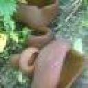
The description of H. rubellus in the link below is perfect with what experienced.
Beiträge von Steve_mt
-
-
Dear friends, I carried out ITS sequence for four specimens from this population and had a 100% result for Xerocomus rubellus MN685116 for three and 99.87 for the other.
Now I am wondering at large why non of the specimens produced the characteristic red dots at the base of the stipe and instead it formed a reddening under the cap. I guess I should trust the result but hmmm ?!???
What do you think ?
I can share the sequence results.
-
I am back with this one and I got the result from ITS sequencing as Psathyrella panaeoloides. As the name suggests this Psathyrella look like a Panaeolus so it is a consolation for me for opting to be a Panaeolus. Schupfnudel you were right on suggesting Psathyrella!!! I was confused between the two but stipe was a bit flexible and the deposits on the cheilos were so much misleading (and the white edged lamellae too)!
Comparing with this excellent report:
Psathyrella-panaeoloidesDaD.pdf
My collection have those ' rectangular ' spores, in the right sizes and some comments make reference to cheilocystidia having deposits on the top. So we are good!
-
Hi Andreas, Thank you for keeping this in mind and I also have not forgotten this fungs on two aspects.
1. If you need to have a voucher specimen (dry) just to have a look, I can send it. I will sent to Alvarado soon. I suspect it is a new species, I posted on MEditerranean fungi FB and they could not come with good suggestions (somewhat trending on Clavaria).
2. I went to check again the micro of my specimens stored in the fridge and stubbornly no reproduction. The basidiole-like hyphae could indeed be immature asci after all.
So this fungus will go to Spain with my next batch.
Let me know if you need a specimen (Message privately yr address or on info@maltawildplats.com)
-
I skimmed over the paper - very, very interesting and the pieces are fitting nicely together. This fungus seems quite close to Hypoxylon or Biscogniauxia anamorph, which then becomes a black stroma - the sexual state - on which the Cosmospora grows on (assumingly on the dead stroma, but not sure). Today I will investigate at high power the condiophores. Biscogniauxia forms black stroma on bark so we are getting close!
-
Well done guys - I come back in the next competition
My favourite one was the Lepista which came 2nd for its subtle colors, angle and soft focus in the border.
-
Thank you xxxx
I will investigate and let you know. To tell you the whole story, I think when its life is over it deposits as a black layer on the bark, on which an interesting Cosmospora s.l. sp. (cf flavoviridis) later grows upon. I really wish to solve this puzzle so many thanks for your guidance.
Nectria cinnabarina s.l. (or something else) - Forum ASCOFrance
-
Hi Andreas, thanks for yr help. I have sent material to Alvarado and I can send this to him too. However if u wish to have a sample to examine under yr lens, I can send you one fruiting body. When they dry they are quite small and shrivelled so hope they rehydrate well. Let me know (send address). The samples in the fridge are also healthy and will check their maturity tmrw
-
Photos in situ....
-
Hi, here I am writing again about another strange fungus growing on the bark of a dead tree that I suspect is Ficus carica. First seen, it looks like a dry coral mushroom with a pale beige-ink color. It grows in tufts and then it apparently blackens out. Under the microscope, it turned out to be a synematous microfungus composed of sterile structural core of thick entangled hyphae, mustard-brown to curry-brown in color but dull. This becomes black in KOH. The outer layer is a thick dense coat of penicillate hyphae, with branches arranged in whorls and each subbranch seems to have a small head of phialides each producing one conidiospore. Conidiosore bean-shaped. This layer is pale mauve (almost greyish) and turns dirty green in KOH. The conidiospores are cream-straw color and become mauve-pink in KOH.
any clues please?
-
Thanks, I try to stay away from FB coz I waste a lot of time, but I think everyone is running out of ideas, and maybe I should post there. However, while the fungus remains in a sterile condition, we can't expect miracles. And I don't feel like going again to the island of Comino for the third time !!
-
Hello Steve,
Water will evaporate out, diffusion is just a matter of time ...
But anyway you can't propably open the dish slowly enough, without creating turbulances, transporting spores inside.
Surely won't work. Sorry, silly idea of me.
Best regards, Martin
It's Ok Martin don't worry, we share opinions without any regret! I was also a bit silly to think it may take 3-4 days to evaporate, but it was quicker (at 40C). Thanks for your thoughts!
-
I managed to dry it with the filter paper method after sterelizing it in the microwave at 440W for 5 minutes. Maybe I am making lot of fuss for nothing because the agar dried in c. 30hrs and that is too short time for any serious contamination.
Now what about storage? I have in mind to l cut the agar around the colony wrap it in aluminium foil and put it in close dry environment, perhaps tupperware container with some silica gel. I am sure insects will feast on the agar if I leave it in a box. I have lot of silverfish in my house recently (probably they are attracted to fingi)
-
Hi steve,
Firts of all, I haven't done this before, but why don't you turn yout dish upside-down, remove the cover and dry it like this?
Contaminating spores won't move upwards, I think.
Regards, Martin
That way the water can't evaporate out
-
Under the microscope, there is more or less the same picture of before, but what we assume that are basidioles are more wriggly (curved/flexuose).
-
Hello... Sorry for letting you wait.
Here are some close-up images of the fruiting bodies, 6 days after the first encounter. The have not changed much. They seem to have more a coral-fungus approach then that of a jelly baby fungus. The flesh is soft and breaks away easily, but decivesly not gelatinous. I have dried some more specimens and placed few in the fridge. Not the heavy pruinosity near the stipe.
-
Found them. 3 populations. Here are quick mobile photos 😍
I take some pics with slr. They did not change that much. Cu
-
We are going for it. Hope we find it in goid shape 🙏😊
-
You hit the nail on its head re last fungus. I see if I can do more re species level! Interesting!
Thanks!
-
But spores, bacteria and living matter that have water inside so the microwave radiation "should" destroy them, at least theoretically. The question is if the rating of 440W is enough, because higher ratings damage plastic, wood/paper, rubber and arcs with metals etc.
Microwave sterilization - PubMed
Effect of microwave radiation on Bacillus subtilis spores - PubMed
Yet these claims do not mention the power rating
So I wonder if someone had personal experience.
-
Wish to have confirmation on my ID of Mycena hiemalis for this wood-inhabiting mycena. Saprotrophic on Ceratonia siliqua
-
I know that microwave should kill all living matter. When it comes to plastic or paper or metals, setting at high-power can cause burns and damages to such materials, but at 270W and 330 W (I think also 440W) no damage is created. Do you know if at these ratings for long period (say 10mins) is an effective way of killing bacteria and fungi spores. Sometimes I re-use petridishes coz they are expensive.
-
Guys, for the last 4-5 years I have been trying to identify a Talaromyces that specifically grow on seeds of Washingtonia.
You can follow and see the pics here:
https://mushroomobserver.org/262509
To sequence it, I have to isolate it on media, which I managed. Now I need to dry it. I have a home made incubator with temperature control (now set at 40C). My plan is to put the petri dish in it but the water moisture evaporating out will condense on the lid and back into the media. I thought of uncovering it but I am afraid it gets contaminated leaving it uncovered for a day. Ideas:
Drilling the lid with small hole / holes (less chance of contamination)
Place a sterile filter paper instead of the lid.
Sprinkle over sulfur to inhibit growth of any fungi (just a weird idea!).
Put petri dish it in a sterile, close tupperware container with the lid open over a bed of dry silica gel
How should I dry it without contaminating it.
-
The specimens started to mold slightly :-(. Yesterday I did not put them in the fridge, so as to accelerate the development process. The best specimen has been dried yesterday so safe from contaminants. Today, I thought, dass die surface was more pruinose ... maybe they make it to sporulate before parasite takes them out.
Andreas, KOH on surface - no color change. It wasn't gelatinous (mucoid or watery) just soft tissue
Yet I found some other strange finger-like organism growing close to them from the soil. Maybe Ceratiomyxa ?!. I may find time to examine under the microscope!
-
Dear Pablo,
Thank you for your reply and teaching. I could clearly make up two types of hyphae and maybe the third one, the binding hyphae (not sure) as shown in the new image here (Sorry for the crap ones before, I was super tired!)
Thanks a lot for yr help
LG
Stephen