


Specimens found, rather workable, I follow up. I am thinking Pluteus nanus for the small size and already found it in three locations in Gozo (kinda frequent).
Beiträge von Steve_mt
-
-
-
I still have the specimen. Is it of further use to recheck something?
-
Hre it is photographed on narrow 'twigs'
banco de setas coprinopsis goudensis coprinus goudensis ulje | www.bancodesetas.es
-
It is a very curious Coprinopsis. Many things match for goudensis too, although admittingly the velar hyphae do look thick (but how important is this?). Then the habitat for goudensis is lignicolous and in my notes, I have written dry debris of herbaceous plants, but I have not checked very carefully the debris and maybe it was mixed with some slender twigs?!?! I can see parallel ridges along the substrate indicating herbaceous stem that dried out but I don't know which and again it is a bit (very bit!) thick and lignicolous. Ajjj!
-
These photos are from an old collection and I don't remember much about it, or if I have a specimen, but this photo shows clearly spores in the range of 8.5-9.5um length and pleuro- (or cheilo-) cystidia that are broad and short. I hope there weren't two or more specimens of different species close to each other and have a mixture. The substrate was annual herbaceous plants (maybe grasses?) that dried out in summer and then this fungus grew after the autumn rain.
-
 Alles anzeigen
Hello,
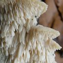
even more interesting to me would be the ascomycete on the wood (?) beneath the Pluerotus

I don't remember to have seen such a Pleurotus, but on Malta there might be Pleurotus species not occurring in the rest of Europe.
all the best,
Andreas
Ooohhh, such a pity because I sent my sample in Eygpt to a mycologist to identify the species but apart he has not carried out any investigation, he kept (or lost?) the samples and never replied to me when asked to send my sample back. I check if I have something left maybe I can send it to Spain. I presume if I find a dry specimen it would not help to carry out further investigations (micro or chemical)?
-
 Alles anzeigen
Hello!
Yes, microscopically that looks like a Pleurotus species. But wich one could be difficult, because the microscopic features of all those species from the ostreatus - group look nearly the same. The substrate would be interesting, was there wood beneath the soil? Because if not, and maybe the fungus grows on roots of smaller plants, that could be interesting.
LG; Pablo.
The substrate was wooden chips and some gravel that was used as animal bedding (and hence it might have some excreta) dumped on soil in an olive grove. There was some burning too but it is probably not relevant. So We could safely exclude roots of smaller plants. I wonder if we can advance further or stop as P. ostreatus s.l. ?
-
 Alles anzeigen
Hi,
definitely not a Pluteus.
 I think Pleurotus sp. is correct.
best regards
Stefan
Yes, I wanted to write Pleurotus like in the title.
-
Sorry for sending you crap data and non-representative photos here. I try to retrieve a specimen from the collection box, keeping my finger crossed the specimens are still intact and will follow up. I am quite sure the gills are free - I add few more photos. Its habit and colours (overall) reminds me in Pluteus nanus
-
Thank you Andreas and Mollisia for helping out.
Host / Habitat: Damp and old stems of dead herbaceous plants
Pleurocystidia size (um):
84.83 x 43.75
86.12 x 48.97
94.08 x 47.90
59.24 x 33.06
58.74 x 35.24
83.79 x 47.61
90.78 x 45.38
Location Gozo (Matlese Islands)
-
Pluteus sp. ? Can it be identified from the photos supplied. It was smallish, about 3cm across
-
Gregarious specimens scattered on a clump of woody stalks (I dont know what!) in a damp area. Rather small fungus, gills free, rim darkens with age. No spore print or micro... hope it is not essential for this one.
-
Dear friends,
I have this Coprinopsis species about 3cm long with partial veil having intertwined filaments with long warts or tubercles projecting out. Cystidia and pleurocystidia balloon shaped.
Spore size:
7.9 [8.7; 9] 9.8 × 5.7 [6.2; 6.4] 6.9 µm
Q = 1.2 [1.4] 1.6; N = 30; C = 95%
Me = 8.9 × 6.3 µm; Qe = 1.4
-
Can you help me with this natural mold forming a dull cinamon / olive-brown mat on the bark of fallen twigs of Olea europaea. The spores are 5um across. There seem to be a verticllaster (whorl) of 3 to 6 phiallides with a swollen head with minute pegs each giving rise to a spore. If further measurments are required, I can carry them out. Thanks!
-
From where they extract genetic matetial to sequence? Do u know? Spores not suitable i guess.
-
I am writing a report on this fungus and I wonder which taxon I should use - ephippium (1841) or sublicia (1799)?
Maybe this recent paper by Skrude and Scumaker (Dec 2020) : ( https://www.researchgate.net/publication/340431565_The_genera_Helvella_and_Dissingia_Ascomycota_Pezizomycetes_in_Europe_-_Notes_on_species_from_Spain ) using the taxon sublicia is the answer.
-
Very nice - I love slime moulds so much, I think the only group which still mostly rely on morphology and only occasionally on genetic sequencing. Very nice specimen there - bright amber-rust - love it.
-
DOWNLOADED xxx
Many thanks, Sir Nobi - They will be helpful and makes my Digital Library a bit more interesting!
-
I am thinking that we should be around Cystolepiota for that fringe and flocculose cap.
-
Hello, I am back on track to identify this small mushroom. Gills are free (or slightly adnexed). Stipe bruises a little (?); cap floccose and appendiculate
-
Alles anzeigen

Hi, can someone help me to find this document:
Synoptic Keys to the Sclerotiniaceae & Rutstroemiaceae in Nordic Countries [Norway, English]
Look here, Steve.
0 Synoptic keys to the inoperculate stromatic discomycetes in the Nordic countries.pdf
But if it will help you?
If you're interested in a special genus, ask me.
There are some more documents from Schumacher & Holst-Jensen, which I can send.
Regards, Nobi
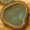
I am interested in the genus Sclerotinia - I only found old literature regarding morpghological identification of three related species. I believe my specimen is S. sclerotiorium
-
Good afternoon dear friends! I wish to thank you for your help. I finished my writeup about this species and I agree that all species in the genus have a Sclerotium and turns out that it is very diagnostic to distinguish the species. I had to base my research on old literature:
- Hall, R. & Boland, G.J. (1994). Index of plant hosts of Sclerotinia sclerotiorum. Canadian Journal of Plant Pathology 16:93–108. doi:10.1080/07060669409500766
- Willets, H.J. & Wong, J. A.-L. (1980). The Biology of Sclerotina sclerotiorum, S. trifoliorum and S. minor with emphasis on specific nomenclature. The Botanical Review 46(2):101–165
- Kohn, L.M. (1979). A monographic revision of the genus Sclerotinia. Mycotaxon 9: 365–444
Three related species are S. trifoliorium and S. minor but my specimen corresponds to S. sclerotiorium . Interestingly it was associated to Chlorophyton comosum, the only herbaceous weed that was present on site, and then a tree of Bay Laurel.
-
Hi, can someone help me to find this document:
Synoptic Keys to the Sclerotiniaceae & Rutstroemiaceae in Nordic Countries [Norway, English]
The link for the file is broken on that site.
I.s the black sclerotium typical/distinctive for the species? (seeing its epithet is sclerotiorium)
Thanks
-
Thank u Stefan for confirming 🤗