Hortiboletus rubellus s
Dwarf version
Troll version
Hortiboletus rubellus s
Dwarf version
Troll version
![]()
![]()
![]()
![]()
![]()
![]() - IT IS THAT ONE!!!!!! 100%
- IT IS THAT ONE!!!!!! 100%
I can double check the spores with x1000 / heated cotton blue, but it matches in all other details.
https://www.myko.cz/myko-atlas/Pholiotina-dasypus/
Thank you so much Raphael - I can sleep well tonight ![]()
Can you msg me your full name for acknowledgements purposes in my paper?
Agreed, I am convinced it is - spores and cheilocystidia matches very well with this species (group). However, I can see that there are minute but constant morphological traits within the complex.
Anyway, doubts cleared - Candolleomyces (Psathyrella) candolleanus... Thanks!
I am getting inclined over Tubaria furfuracea aggr. Despite the adnexed gills are not typical for this species (but I saw one collection which was like this example on an Italian forum) the cheilocystidia are subcapitate (without a constricted neck) as in Tubaria, while the COnocybe I checked (herbarum, brunnea) have distinctly capitate cheilos. Yet it may still be some other Conocybe or another genus that I am not aware about. The spores match nicely where in Conocybe spp I checked, they are a bit more ellipsoid rather than longitudinally asymmetrical (almond shape) as in this case. I dont know who agrees with my assumptions - difficult puzzle!
The above also makes reference to a darking stipe towards the base which is not exhibited in the studied specimens! (but then Tubaria's stipes are seldom whitish and more robust grrrrrr!!!!!!)
I considered Galerina clavata too but this has larger spores >11um and cheilos very capitate
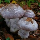
To confirm that all these specimens within a common population growing close to a trunk of Salix alba are all Psathyrella candolleana. A small doubt is arising because the gills and spore print is (maybe) too much chocolate brown and usually, it is lighter and has that bit of a violet-mauve hue.
Spores
(6.7) 7.1 - 8.4 (8.7) × (3.7) 4.3 - 5.1 (5.5)
Q = (1.5) 1.55 - 1.8 (1.9) ; N=26
Me = 7.8 × 4.7 ; Qe = 1.7 ; Ve = 90
Another option is Psathyrella bivelata, said to have dark tufts of veilar remnants on the pileus
Alcune specie interessanti del Genere Psathyrella
Thanks for your opinions.
Hello to everyone. The rain in Malta is finally back after a delay of 6 weeks and the mushrooms start to appear. I am struggling with a Conocybe (or a Tubaria/Bolbitius?) and wonder if I can find some help on this wonderful platform.
Three fruiting bodies from leaf litter of Olea Europaea (and Ficus carica) or jus from peaty soil below. Spore print tobacco brown, pileus wet, toffee-brown with evident radial 'ribs'
Spores, almond shape, with a large/wide apiculum
7.8) 8.1 - 9.3 (10.7) × (4.3) 4.6 - 5.1 (5.4) µm
Q = (1.6) 1.7 - 2 (2.1) ; N=37
V = (81) 89 - 124 (166) µm3
Me = 8.8 × 4.8 µm ; Qe = 1.8 ; Ve = 108 µm3
Cheilocystidia capitate with a strange banded staining in Congo red (apical part stains stronger). Pleurocystidia possibly absent. Basidia stout, potato-shaped or sack-shaped with 4 tiny sterigmata.
I don't have keys of this genus, although some ideas I made are C. herbarum or C. juniana. C. brunnea (or maybe Tubaria hiemalis)
Thanks guys, then it is easy quite easy to distinguish these two Coprinellus and I shall learn these characters for future reference. No setae observed on the stipe.
Great!
Thanks again.
 Alles anzeigen
Hello, my dears!
Since after an hour of mushroom presentation my head was spinning with all the interesting mushrooms and their mostly scientific names, I had to draw something...
Warm greetings,
Tuppy
How did you draw the sky/clouds so good!
Hi I found one of the first clump of macrofungi after the first rains a week ago and it corresponds to the usual Coprinellus but I have to decide between C. micaceus or C. saccharinus. What clues do you have for me? I have a key but I want perhaps have fresh ideas on this platform. Many thanks
Many thanks for confirming G. resinaceum - First time I am seeing this species. I will go again to take more photos and maybe some spores.
KaMaMa, last photo with mouldy patterns is really cool!
Yes, autumn is back to our Islands, although it rained once about 10 days ago and no more for the next week. But you should see me a bit around again!
I've today spotted a developing Ganoderma sp. on the trunk of a n oak tree, here in Gozo, Mal ta. Do you know what is the species at this early/immature stage please? I have thought about G. lucidum or G. resinaceum.
Thanks and reagrds
Stephen
I just discovered this cute tiny fungus (not really mould or microfungus so hope here is a good place to post) and wish to share with you. It was growing on several pinnae (leaves) of Phoenix cf dactylifera.
Now I want to identify this Aspergillus which was growing on the Parsley flowers and fruit together with the Erysiphe
Part 2: How did I manage to isolate the Aspergillus!
I took a the plate which had a dominant colonization of aspergillus, remove the lid, and tapped gently over a plate of innoculated Oat Agar. Theoretically, only spores of Aspergillus should drop.
Result: Success ![]() (plus a tiny contaminant of A. niger! - very persistent )
(plus a tiny contaminant of A. niger! - very persistent )
-)
So here we have a situation of buy one and get one free. I cultured the microfungus by touching the infected fruits on petri dishes, and what we have: two microfungi - The Erysiphe with many white unornamented Chasmotheca intermixed with an Aspergillus species forming beautiful cyan / sky blue conidiophores.
I tried to isolate the two by careful inoculation by I got them both growing every time
Agree, I just find it strange the growth is only manifested on the inflorescence!
Thanks for replying
Here is a link re disease of parsley (general info)
http://ausvegvic.com.au/pdf/Parsley_Disease_Handbook_2006.pdf
Is it normal for this species
to grow only on the fruit?
Thanks, I check the prices, and if they deliver to M Alta coz These are the top Lab suppliers and I do not need Grade A stuff...
Thanks mate
Is there a particular species of Erysiphe that grows specifically on the fruit and flowers of Petroselinum crispum (not on the leaves). The leaves are intact. Logic says E. heraclei but not so sure since of its specificity on the reproductive parts of the plant.
I am trying to order it. 80Eur is worthed (NHBS is ca. 150 Eur!)
See if ths helps ![]()
 Alles anzeigen
Hi Steve
You can use different Genus, which are in general mycorrhizal
ground floor Cortinarius, Inocybe, Hebeoma, Russula, Lactarius, Amanita, most of Boletales
...
As I know for aforestation
Leveroma is a good choice
BR
Uwe
Thanks UWE, is it as simple as that ![]() ??
??
Many thanks for all your help, and sorry for my late reply.
You are suggesting a number of genera, but how do you know that they are (from experience and articles probably) but I would love to buy a book (or paper) listing numerous examples and which trees they are suitable for. From experience, I know Suillus collinitus is mycorrhizal with Pinus... but its would be great if I can have some reference book.
E.g.
(...but for Europe?)