


Marcel , thank you so much!
I had these but I think I had an older version (2017).
Good day
Marcel , thank you so much!
I had these but I think I had an older version (2017).
Good day
Some reference links
C. cinereus: https://www.grzyby.pl/coprinus…ljee/species/cinereus.htm
C. macrocephalus: https://www.grzyby.pl/coprinus…ljee/species/macrocep.htm
C. lagopus: https://www.grzyby.pl/coprinus…ljee/species/lagopus_.htm
Key: Sect. Lanulati: https://www.grzyby.pl/coprinus…ee/species/Coprinus.htm#J
Either macrocephalus and lagopus....
 Alles anzeigen
Hello, Manfred!
It is definitely an impressive sight.

Even if the distinction here is rather trivial: Andreas also has a real >Ink Cap Key< on his website , which can do much more than the key in the large mushrooms.
LG, Pablo.
The link is not available, can it be found somewhere else online ?
LG
Steve
May he rest in Peace ![]() He was so much helpful and seemed a kind, happy go lucky guy.
He was so much helpful and seemed a kind, happy go lucky guy.
Bernd Miggel have a look at some messages re yr post ![]()
Found on a pile of dung (cow most likely) mixed with straw
Spores:
(13.1) 14.2 - 15.9 (16.7) × (8.3) 8.6 - 9.5 (9.8) µm
Q = (1.5) 1.6 - 1.7 (1.8) ; N = 17
Me = 14.9 × 9 µm ; Qe = 1.7
Shape: Elliptical-ovate
Oil bodies: Not present or nor observed
inamyloid, smooth, small germinal pore , rather central
Cutis of rectangular hyphae, sometimes with rounded or attenuated ends (40-)62.4(-80.1) x (12.8-)20.4(28.9)um, hyaline, thin walled, not pigmented
Veil composed of cylindrical hyphae, mostly straight, variable in size but uniformly arranged; (24-)43(-62.4) x (9.3-)13.3(18.3)um
Only a doubt on C. lagopus actually....
Dear all, I have passed from this situation myself that is - C. granulata or C. theleboloides ??? --- and with lots of frustration, sometimes.
I think Moravec (2005) also got into this situation and he dealt by describing var. (or forma) glabra of C. theleboloides , which solve the problem when having a theleboloides with few scanty hairs at the flanks of the apothecium.
P.s. why there is a cross behind Nobi - hope not what I am thinking about ![]()
![]()
![]()
Dear Bernd, I don't know if you are still around, but I was working with a Cheilymenia recently which I identified as C. theleboloides. I am writing to note that the color of the asocarps were initially yellow when young and then they became orange when I revised a 5 days later (and the ground was more dry). The spore striation, habitat and spore size is very important to identify. In Congo red, the striations won't show and in Lactol Cotton blue you need a big effort (or a big microscope) to see it. The hyphoid hairs look like C. Theleboloides except the split hair in yr image (maybe an exception) but the apothecia of my finding were generall larger, ca. up to 8mm.
Hello Matthias,
Sorry for late reply. Actually I am back on track as Coprinellus sp for this finding, rather Psathyrella. Your suggestion fit several characters esp. in the microscopic aspect, but hmmm... the macro comparison is not that very convincing coz my specimens have this tawny (brown) colour whereas the cap of C. hiascens is mouse-grey Although I seen images having a browner colour on the net.
What is so strange with this collection is the lack of deliquescence and the production of few spores observed in all specimens, where the gills remain pale to moderate brown. Coprinellus sp. are usually black and show deliquescence. Another thing is the lack of pileocystidia in the cuticle (or did I miss them ?!? )
I see if I can send it for molecular determination in the future, but it is a very odd finding.
TNX
Hi Marcel, Yes, that makes more sense! I dont know why I neglected this Genus (the animal excreta maybe?!) although the plicate cap (deep furrows) and small size are features of Coprinellus s.l.
hmmm... the more I look at this fungus, the more I am puzzled.
The stem is brown, not much as many Coprinellus which is often beige or white. The spore cover is minimal, indeed the gills are brown not dark-brown / black
I was wondering I I had a mixed collection of two species but it is clearly shown that the lamella are forming the black spores. The gills are then widely spaced apart (most Coprinus are densely packed). The spores however are typically of a Coprinus s.l.
Hello, I am a bit stuck with this finding from the Maltese islands of a Coprinellus sp. on damp soil mixed with farm animal bedding (= rich in nitrites), which gave me an indication around Coprinellus disseminatus, but the micro does not match because the spores are too large. They have a large central pore.
(9.7) 10.1 - 12.5 (13.4) × (5.3) 5.7 - 6.4 (6.6) µm
Q = (1.6) 1.7 - 2 ; N = 21
Me = 10.8 × 5.9 µm; Q = 1.8
Veil elements spherical
Cheilocystida present, not abundant (maybe old specimen) - Lageniform to subutriform, approx. 60 x 20 µm
C. micaceus maybe ?? but not happy, look the gills they are widely spaced (C. micaceus dense)
Mellieħa, Malta, 11-10-2024, under Pinus halepensis in damp soil close to a sandy shore lining brackish water.
Yes it is a possible cause, I can spray some contact cleaner inside the potentiometer.
It can also be in the PSU, current not being delivered at high setting.
Thanks for your opinion !
Regards
Stephen
I have a 40 year old Zeiss AxioLab RE microscope which has been my working horse and faultless for so many years.
Now I have this fault, where when I increase light up to about mark 5 it works fine, but as I increase the power more (to mark 6), the light dims to about level 2. It is not a big issue but maybe it is a known fault and I can inform my electronics friend what needs to be replaced (leaky capacitor in PSU ?)
Thanks
Can you put / write the link again without https://
Stick does not work and the pasted url is 'truncated' so it shows
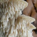
Here, I want to present an interesting early-fruiting species that I only have (for now) macrophotographs because I saw this in a public garden and was not prepared for collection (also images from my mobile) but I am sure it is a Leucocoprinus. Usually these early-fruiting yellowish basidiocarps in artificial soil are L. birnbaumii but this one was different in:
1. Not in compost / peat or pot but more 'outdoor soil'
2. Smaller, thinner, "fragile" - the ring falling off
3. Colours are definitely paler
So then doing further research, I am thinking that I found Leucocoprinus fragilissimus. The only thing is that from many images this species on the net, it seems to have a narrower, tooth-pick like stipe (but some shows similar features).
I can do a collection tmrw, hoping they are not destroyed.
In the image below I slightly enhanced the colours
I always experience poor spore amounts with the small white Phloeomana / Hemimycena ( / Mycena) spp. that I never even think of attempting a spore print. I should do amyloid test from now on, and better observations on the pilleipellis. I look forward this season to study more collections. I think I have some one or two old findings yet undetermined, which I may discuss in this group.
Thank you for your help and correspondence. I will insert a note Det. by Matthias ??? (what surname pls? - send me a private message if you wish to keep it secret from public domain) Btw Happy birthday tomorrow ![]()
Thanks for discussing this and sharing your observations. I agree about not using the extremes of the diopters. Very strange about yr last comment too.
p.s. The link says forbidden ?!
Dear Matthias
I have re-explored my images and I have some further information.
1. Spores:
(7) 8.2 - 9.3 (9.9) × (4.5) 4.55 - 5.1 (5.7) µm
Q = (1.5) 1.51 - 2 ; N = 7
Me = 8.7 × 4.9 µm ; Qe = 1.8
Not very common, based on 7 spores only, but it matches the measurments you suggested
2. Clamp junctions
I might have seen a few in the stipe but not abundant. I marked a few.
3. Inflated Hyphae
You are correct, must be trama / structural hypha under the hymenium
4. Stipe long hairs
Yes, these are very setiform hairs coming out from the stipe. I cant say if at the base or at the apex.
5. Lack of cystidia and pleurocystidia
Agree, absent - they should appear in my slides. I only see few basidia and basidiomes.
The only bad news is that I have not examined and photographed the pileipellis and maybe those you noticed are artefacts. I will study your suggested species shortly. (It looks like it at a glance!). Check some strange structures in one image (below) like a cluster of spores. Some kind of artefact I think.
Matthias, I am hugely indebted to your time and explanation. I will review the images for the details you suggested. Yes, I agree this is a Hemimycena following you line of thought and findings.
Coincidently another member shared a link to a monograph on Hemimycena in Europe by Antonin and Noordeloos (together with other related Mycenoid genera)
I am just working on some Mycena / Hemimycena specimens!!! That's great share. Thanks!!!

hi Steve,
Have you checked amyloidity?
regards Pit
No unfortunately, but my heart tells me it is a member of the Phloeomana group (they are all inamyloid if I remmeber well). Still, I am not convinced it is P. hiemalis (best match) or alba, because there are some considerable mismatches. I discarded the option M. polyadelpha too. Those inflated hyphae are very peculiar!
rare occurrence of caulocystidia
Some time ago I have found a cluster of small Mycena growing on decaying bramble sticks (Rubus ulmifolius) having a purely white and slightly transparent colour (esp. the stipe), fading a bit into beige when old. Cap 7-12 mm wide with non-numerous, spaced (not crowded) lamellae, horizontally-arched not particularly decurrent. Basidia 4-sporous (sometimes 2). There were swollen hyphae from sub-globose to broadly cylindrical, sometimes with a short foot (sphaeropedunculate) which I think were cheilo or/and pleuro cystidia (40-90 x 25-50 um). Spores 8-10 x 4.5-6 um (Q 1.5-1.9) ovate-elliptical to moderately broadly ovate, very rare to find in slides. I didn't check well the pileipellis. The stipe was more or less glabrous but I think I got a slide showing some caulocystidia.
Do you have any suggestions?
I shortlisted to Hemimycena gracilis, Mycena alba, Mycena pseudolactea, Mycene spiraea