Thanks, will do over the weekend.
spores, cheilo/pleuro, and pileipellis.
Thanks, will do over the weekend.
spores, cheilo/pleuro, and pileipellis.
Thank you, we have the same thoughts on this. I'll check the micro shortly. I was wondering if it is too small for L. subincarnata?
Thanks for replying
I have another Lepiota to show you. No microscopy for the time-being but specimen are being dried. Three specimens growing under oak tree (Quercus ilex) although Pine trees are also present not far. Specimen small, around 3cm tall with a cap about 2cm. Ring present but deteriorated with time. From my understanding, the stipe darkens to reddish brown with age that at one point when at home, I thought I might have gathered two different species, but on seeing photos in situ I believe they are all the same species. Margin pleated. Very elegant the tufts of veilar remains, toffee-brown in colour. Your opinions are welcomed on how to help to get an identification, unless molecular sequencein is the only way forward. Thanks!
Hello, rainy and cold season started in Malta and the mushrooms are punctual.
I was lucky to find something quite rare (for my eyes!) a small Lepiota with nice pattern of veilar remnants on the cap. I have dried a specimen for later microscopy, but at the moment I wish to have some clues. The fungus was about 2.5 cm tall and a cap of about 2 cm across. Scent indistinct or faint. It was growing in a mixed aforested area in a valley (sem-riparian) and the two trees present were Quercus ilex and Pinus halepensis. I have got an idea of L. subincarnata or because it is small, L. echinella.
I can carry microscopy after 5 days because I am away from my home Lab.
It has been decided at this hour during a meeting by JONEF members and we updated the deadline during this meeting to 10th Dec. It will be shown online soon, too.
Tnx
Dear myco friends,
I am part of a project called Jonef which is building a report for the conservation and protection of fungi based on two questionnaires.
Info on the project can be found here
Project JoNeF – Joint Network for wild Fungi | Network per la diversità micologica
The direct links for these questions are found here.
PART 1
Füllen Sie bitte dieses Formular aus
PART 2
Ausfüllen | JoNeF Questionnaire-base Survey (Part II)
Deadline 10 December.
It is an easy online data input.
Your participation is welcomed and important especially because we lack data from germany, poland (and spain).
Thank you
This is Biscogniauxia mediterranea on dead Quercus ilex tree.
Alles anzeigenHello Steve,
so far I think the new system is even better than the old one!
But I understand what you mean.
By the way: Fuligo was not placed with Diderma but Mucilago with Didymium, but I could well imagine that Fuligo will soon belong to Physarum.
Kind regards
Noah
Yes, sorry - bad memory - I meant that combination -> Mucilago into Didymium
For a long time the study of myxomycetes was my favourite because the classification and keys and genera were quite straightforward and there was not much interference from genetic disruptions. Hope that more or less, things in slime moulds remain like this. Having Fulgido absorbed in Diderma is acceptable but let us see what happens in Badhamia and Physarum and if the basics we learnt have to be erased with new molecular concepts that not always make straight sense to a sensual brain!
![]()
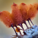
I have found this cluster of myxocarps aggregated in tight groups (but never in plasmidiocarps) on a decaying legume (fruit) of a carob tree. After sitting in the waiting list for months, today I decided to study this slime mould.
The myxocarps are about 0.5-0.9 mm across, seem spherical but a lateral view shows they have a conical blackish base and sometimes a cream-mustard pseudostipe, making some of the fruiting bodies have a pear-shape structure. Interestingly the lime on the peridium is not thick, rather vein-line with nodules scattered here and there and overall, the peridium is moderately irridiscent esp. in artificial (LED) light.
Now comes the hardest part of me - Badhamia or Physarum type of capillitium?????
Essentially, the structure is made of columnar lime with various Y-shaped particles but also with fine, hyaline threads from them. For me, it is more a Badhamoid capillitium, but not sure.
The spores are globular, spinulose-punctate with very tiny dot-like projections rather than warts of spines, evenly distributed without dark or pale patches or lines or irregular patches whatsoever. They measure (8.5)9.0-10.0(10.5) um across, rather homogenous.
In the end, I concluded it is Badhamia foliicola (second preference a Physarum cinerea) but I wait confirmations after I assign this name to the species.
Thank you
yes, truly beautiful species both as a singular myxocarp as you well explained, and also in groups, with those quite equidistant spaces between sporocarps making everything very pleasing to the eye.
Thank you for the update, something we need to get familiar with although the changes do not seem as drastic as in Fungi and to a less extent plants (although placing some snapdragons with same family of plantains is still something I am still digesting slowly)
About two weeks ago, I came across a strange coal-like fungus on the bark of a dead, unidentified tree, which had the texture and looks of a Daldinia, but it was flattened so I could not assign it as the two species known in Malta - D. concentric and D. raimundii. These have distinct semi-spherical or cushion-like fruiting bodies, with concentric rings, hence to exclude, but not sure if other Daldinia species that are flattened. I have now considered Biscogniauxia sp. but not sure which sp would be. I can revisit the population for microscopy and further examinations. The tree is not beech, maybe Quercus
Dear Raphael,
Many thanks for your feedback. Tomorrow I send the main corresponding author an email and see what I get. Despite DNA analysis via Blast and GenBank, it is not always assured your best-matched sequence has the correct name, hence studies like those you mentioned come useful. Thanks again!
Alles anzeigenHello
Very interesting Lepiota in such small cases, I would also bring following Option in the inner choice -> Lepiota pseudolilacea
In the fourth picture the color is more black, are you Show two different types?
Best regards Andy
Edit, Chris was faster 😉
Hello Andy, thank you for your suggestion too. I appreciate
The fourth pic is the same specimen but not in natural light but under the stereomicroscope with badly assigned White Balance. I played a bit with the contrast too so ignore the colours in the 4th pic. Yet when i dried it, the colours darkened.
Alles anzeigen
Thank you for your opinion, I was unaware about L. pseudolilacea (does it grow in this habitat too?). I admit that it resembles it a lot, although I would love to know the major distinction between the two sister species. I see if I can find some literature on my behalf !
Hi, I have sequenced some specimens of Pluteus, and would like to know if someone is working on this Genus and if my DNA sequences from Malta would be handy. I have sequenced this population with slightly yellowish stipe, and the LAB suggested Pluteus cf nanus. I am not sure if it is this species if it the name was not correct when the author of that sequence made a good or bad identification.
Anyway, I would love to hear your opinion on this Pluteus sp.
LG
Steve
This small Lepiota sp. was found on moss and grassy soil in a damp site over calcareous limestone. It was not in a very good state but I could see a brown-purplish ring which alerted me on L. lilacea. On age this darkened to blackish-purple-brown.
I have some microscopic data too if required. Spores scarce, could not find them
Dear friends, I need your opinion on this crust Polypore I found on the wet bark of Ceratonia siliqua.
It is a white polypore which seems to be accompanied by its amorphous stage as grayish - olive green colonies at the perimeter of the crust. I include several photos of the teleomorph and anamorph, both macro and micro.
NaOH gave a green color change on the hymenium.
Spores bean-shaped, about 5um long
Measurements:
Basidiospores
(3.9) 4.2 - 5.5 (5.8) × 2.2 - 2.7 (3) µm
Q = (1.6) 1.8 - 2.1 (2.2) ; N = 18
V = (10) 11 - 22 (23) µm3
Me = 4.9 × 2.5 µm; Qe = 2 ; Ve = 16 µm3
Cystidia/Basidioles - 12.0-17.0um x 4.0-5.5um
Ascospores:
(12.7) 13.9 - 17.2 (18.6) x (9.1) 9.3- 12 (15.1) µm
Q = (1.1) 1.3 - 1.7 (1.9) ; N = 42
V = (546) 680 - 1210 (1985) µm3
Me = 15.7 × 10.4 µm; Qe = 1.5 ; Ve = 910 µm3
Basidia
19.10 x 5.4um (Sterigma = 2um)
20.5 x 5.7 (Sterigma = 3um)
I always have problems with crust polypores but Ropia and Ceriporia crossed my mind.
Regards
Stephen
Amyloporia xantha in its early stages. Sorry for taking so long to come back on this
Hello everyone, I found this beautiful population of earthballs (Scleroderma sp.) and studied it for Identification. The peridium had a nice pattern of dark brownish patterns (referred as leopard skin) on a cream/light yellow background, later becoming beige, and on cross section it was egg-shell thin (less then 1mm). Basically, we are in the S. verrucosum / S. areolatum area, and since the spores were between 7.5 and 9.0 um (Average 8.23 um) with numerous straight to curved spines about 1um long, I think it can be identified as S. verrucosum
Hello I have met this scleroderma and I assume it is S. verrucosum from its eggshell-thin peridium, leopard pattern on a faint yellowish-cream background, small size (2-4cm across). What does not fit is the absence of a pseudostipe, and the spores are a bit on the smaller size (7.5-9.5 um, averaging 8.3 um).
Some years ago I had found a similar Hygrocybe (under olive trees and Pine trees) and upon researching, I had read that if it is not slimy, it would be the Hygrocybe conica s.l. whereas if slimy it would be attributed to H. singerii. Now I wanted to know from your experience how much singerii are actually recorded from Europe and if that claim (slimey=singerii) applies for Europe. Tnx in advance!
