


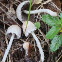
Very small fruting body (1cm) , not sure if it is the normal size, cap very fibrous - Inocybe sp. ?
Sample collected
Very small fruting body (1cm) , not sure if it is the normal size, cap very fibrous - Inocybe sp. ?
Sample collected
Hi, I found this black cup fungus (Ascomycete) and I considered Smardea and Pseudoplectania but now I am fixed on a dark-coloured Peziza sp. This population growing in mossy ground under Pinus halepensis in a forest path, in calcareous soil. In sunlight, the ascocarp has a purplish-brown tinge. I am showing this collection from photos I have taken in the field. Further examination in the Lab follows shortly.
I will produce images of spores, cheilocystidia (and pleuro) and pileipellis today. If other features are required I let you know. (The english translation of the fungus you suggested is "Cadaver"!!!)
Thank you so much for these helpful notes. I was away from mycology for two days but today/tmrw I should do some investigations. The fruiting body has a hint of blue. The stick was found around and probably is of Pinus halepensis. I consult Bernnichia's book . I keep you posted. tnx.
Can this small pore-bearing fungus (Polyporales ), approx 1 cm wide, be identified. The pores are very minute, barely visible with the naked eye. They were attached to a stick and I think they were facing down (hence not resupinate as the pic may indicate).
Hello, I found this nice species of Inocybe growing from leaf litter of Pinus halepensis and I think (not sure) to correspond to Inocybe rimosa (=I. fastigata). The colour, cracked cap and fluffy stipe corresponds. Wonder if you have different opinions or maybe there are other newly described species within the rimosa group.
LG Steve
The images are amazing. Do u have the monograph ? If not I can send
Thanks, Clavaria for the info.
I reworked the spores and the sizes are those below, making it a clear D. coprophila (spore size = mean 12.4 x 7.6)
(10.4) 11.8 - 13.2 (14.1) × (6.2) 6.9 - 8.2 (9.3) err
Q = (1.5) 1.51 - 1.8 (1.9) ; N=31
Me = 12.4 × 7.6 err ; Qe = 1.6 ; Ve = 381 err3
 Alles anzeigen
Hello!

Clitocybe it is. I think it can remain within the variability of C. hobsonii!
The real name is "Clitopilus", not Clitocybe.
http://www.pilzflora-ehingen.d…ora/arthtml/chobsonii.php
VG Ingo W
Dear Ingo,
Yes I got tired yesterday - I corrected the names. I agree re hobsonii, yesterday night I found the differences between the two, mainly the spore length with hobsonii being: 7.5-8.5 and daamsii : 8.0-11.5 um. I really a merry-g0-round here but it is nice to learn. i will never forget the vertical ridges now ![]()
Thanks to all - I appreciate a lot
LG
Steve
I abandoned C. hobsonii because of its smaller spore size. Also, the robust apiculum is something which mislead me. But yes, I agree about the longitudinal shadows of the spores (see a better image here!). Moreover, I went to check for the Cheilocystidia and could not find any -I think what I am seeing are basidioles. If I remember what I read in the afternoon, lack of cheilocystidia is a Clitocybe character.
Clitocybe it is. I think it can remain within the variability of C. hobsonii!
Further research and applying the key on European species, I have landed on Crepidotus subverrucisporus , typically on calcareous habitats
https://www.mykoweb.com/Crepidotus/species/Crepidotus_subverrucisporus_subverrucisporus.html
C. variabilis has smaller spores (Sp 5.5-7.5 x 2.5-4 μm), and more narrow - subcylindrical
C. cesatti has more broad (subglobose) spores (Q. <1.4)
C. luteolus has yellow tinge (at the less)
C. lundellii* is closely related but it is said to have brownish colours when dry and not calcareous
So at the moment, this is Crepidotus subverrucisporus or C. lundellii - I guess I have to re-examine the cheilocystidia?
C. lundellii: https://www.hlasek.com/crepidotus_lundellii1en.html
* One of its synonyms is Crepidotus inhonestus !lol! - why this fungus is dishonest haha!
I have given a quick check at the spores and I think you are right about Crepidotus - the spores are too large for C. hobsonii, and their shape, a bit like a lemon due to a distinct-swollen apiculus is also Crepidotus rather Clitocybe.
Spores:
(7) 7.1 - 8.3 (8.9) × (4) 4.1 - 4.8 (5.2) µm
Q = (1.5) 1.52 - 1.8 (2) ; N = 12
V = 61 - 98 (110) µm3
Me = 7.6 × 4.5 µm ; Qe = 1.7 ; Ve = 81 µm3
I think you are correct (or Botryotinia) but it must be a species that does not have a branched conidiophore. I always associated Botrytis with a branched conidiophore (that is I have always seen B. cineraria). I see what I find in this genus with large spores. B. elliptica for example!.
Thanks for the moment !
 Alles anzeigen
Hi Steve,
compare with Botrytis sp.
best regards,
Thorben
OMG there is also subcoprophila! How to tell the difference between the two? I have some microscopic data at hand, such as the spores.
Deconica coprophila on horse dung pellets - are all four the same species ? Maybe the third one is something else ?
Thanks
I forgot to add that I managed to get a pure colony on Sabourad Agar. It formed a white or light grey mycelium without quick fruiting bodies. These were formed after 1 week when the mycelium covered the whole plated and they were formed at the walls of the petri dish (drier regions ?), somewhat not that black and shorter. Now I have inoculated OAT, PDA and CZapek to see if it helps. Really wish to learn this microfungus. Chloridium is close but spores are larger 10-17 um long, 8-11 um wide
Growing on the pulp of a fallen fruit of Hawthorn (Crataegus monogyna) was a dark mycelium with white heads which took my curiosity to investigate. Under the microscope I could see a simple, dark-coloured (blackish-brown) septate conidiophore (somewhat paler at the tips or undeveloped ones) with a small head-like cluster of hyaline-white conidiospores. These are liberated readily in water or soap water, so I suspect that there is some slime involved. However, at the location where the conidia are present are 3-6 small, flattened plugs projecting out onto which clusters of spores are attached. The spores are subglobose to oval, variable size, possible not hyaline but opaque since spore contents (eg oil bodies) are not seen.
Hi Thorben, I have found something similar, and I could not pin down the genus, hence posting soon on this forum. Unfortunately, I don't think we have the same organism because mine grows on decaying fruit and the spores are not so hyaline as they dont show the oil bodies.... but is not so different in general. Do you have some key to the genus ?
Thanks for trying to cheer me up! I am a very emotional Brazilian fan, and I need 3-4 days to be back in the mood. Tomorrow I prepare mulled wine to join you in spirit! (...and do some work on this pluteus).
It was nice to see again this tiny orange fungus - Pithya cupressina on fallen leaves of Cupressus sempervirens. (Malta, 10-12-22).
Thank you Harold - later I get grips on Flora Agaracina and dive into the keys. I keep you posted after I recover from mental trauma after my all-time fav. football team got eliminated from the world cup so dramatically - Brazil. ![]()
![]()
![]() I guess many Germans here have passed this already.
I guess many Germans here have passed this already.
I am working on another new basidiomycete, this time a clear pluteus for its free gills (distant from the stipe), rose-light brown spore print and lack of veil, ring and volva. The light lemon-yellow colours of the stipe and the subspherical spores indicates a P. romelii, but the cream with a very faint hue of yellow colour of the pileus is quite off for the species (P. romelli is caramel to cinnamon brown). So here I am discussing this fungus with you.
Basidia oblongish-subclavate, sometimes flexuous, predominantly 2-spored, rarely 3-spored, up to 35um long
Cheilocystidia are large, lageniform to broad utriform (prone to correction!)
Pleurocystidia present, similar to the cheilocystidia, perhaps a bit smaller
Spores subspherical (some looks spherical) to globose-ovate, Me = 7.1 × 6.3 µm; Qe = 1.1; Ve = 152 µm3; 5.9) 6.2 - 8.1 (8.6) × (5.2) 5.7 - 7.1 (7.7) µm (n=33). In lugol's iodine they showed a dextrinoid reaction, while in Cotton Blue (with lactic acid), the spores collapsed inwards.
They were growing from leaf/twig litter of Eucalyptus plantation. I had in mind Pluteus romellii s.l. and Pluteus boudieri P.D. Orton 1960
I am aware by the work of : Holarctic Species in the Pluteus romellii Clade. Five New Species Described and Old Names Reassessed [ https://www.mdpi.com/2309-608X/8/8/773 ] but all their described species do not show this pale pileus. P. boudieri is my best match so far.
Spores in 3%KOH (aq) and glycerol (3:1) at x1000 oil immersion.
In my opinion, the single gap in the spore wall is derived from the apiculus. Saying that images of Ph. nemoralis on the web don't show a distinct germ pore, but often you can see 1 germ pore in ten spores. I have photographed some 200 spores, and I can't say I see any distinct examples. Nevertheless, I stick to the keys by Arnolds (2006) and assign this to Ph. vestita. Also, reading the description of Ph. vestita in Arnolds (2006), I see that it is a good match. Moreover, the notes for Ph. nemoralis mention the close relationship with Ph. velata
.
... on your spore pictures I see something like a indistinct germ pore (callus) on some spores. The spore wall is thinner and/or lighter brown:
Some Pholiotina species have a very indistinct germ pore.
I was gonna write earlier that if my specimen had a germ pore, this would perhaps match Ph. nemoralis. What I can do is to mount the spores at x1000 but photography may not be a state of art ! Yet it seems that there is no germ pore (in these images I took).
I have found Ph. dasypusa a week or two ago (i think I posted on this forum) and it had a different 'feeling' namely the cheilocystidia were more capitate and less broad, the colours of the pileus not so dark (caramel-brown) with less evident margin veil, and more importantly, the stipe flesh was not reddish brown but pale beige. I think we are not 100% happy with vestita for the striations in the pileus right? --- and the colours are a bit too vivid here.
More cheilos with graphic enhancement - lageniform with long slender necks but variable too.