Hi, I was doing a foray in a damp habitat covered in litter of Quercus ilex (Holm oak). There was a large population of Boletales. I have isolated four collections although realistically I think there are less different species and possible there are 2 Xerocomellus / Xerocomus. I have previously recorded Xerocomellus redeuilhii from this site.
1
Small to medium-sized fruiting bodies, scent faint and not distinct, taste mucoid and fungoid, bruising of pores becomes slightly darker (= no colour change), bruising of stipe no colour change either when cut context completely yellow even at the base. Stipe bright yellow without reddening at the base including when cut. Some specimens with cracked pileus showed an interesting red reticulate pattern where cracked. No colour change when pores and stipe are bruised
 IMG_8769.JPG
2. This forms a beautiful pin-shaped reddish fruiting body where it remains small and almost spherical. Taste fungoid and very palatable, scent nil, when cut there is darkening at the base which eventually turns in some streaks of greyish-dull blue at the base. No colour change when pores and stipe are bruised
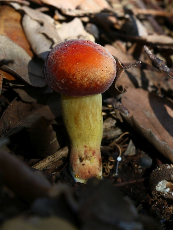
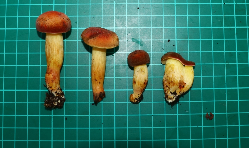
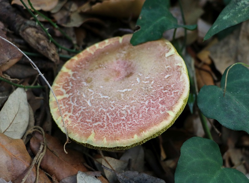
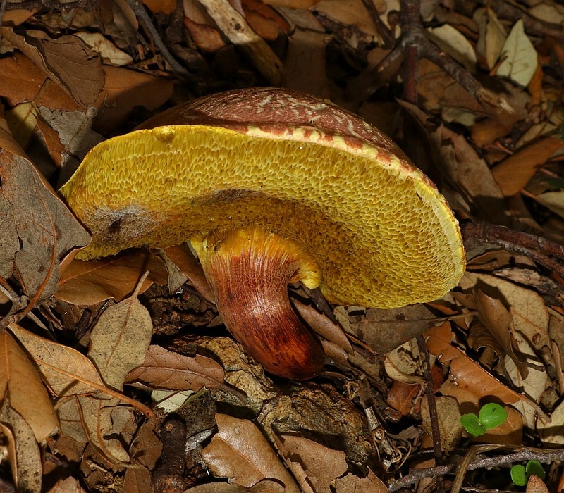
3. The third 'collection' consists of medium-large fruiting bodies, with a pale or light-coloured pileus, tan or peached colour, sometimes forming cracked cuticle with reddish flesh. Stipe red for most of the length (Xerocomellus redeuilhii ?!?)
4. Maybe same as 2 but with flattened pileus
IMG_8789s.jpg
Opinions greatly appreciated. These are all from the same location (100 x 50 m area), calcareous rock or soil in a damp location over leaf litter of Quercus ilex.
Xerocomellus redeuilhii / X. chrysenteron / Hortiboletus rubellus maybe (no.2)