


Coprinopsis cothurnata ?
Coprinopsis cothurnata (Rotfüßiger Schneetintling) – Fundkorb
Coprinopsis filamentifera ??? (I dont think so)
Coprinopsis cothurnata ?
Coprinopsis cothurnata (Rotfüßiger Schneetintling) – Fundkorb
Coprinopsis filamentifera ??? (I dont think so)
This is another specimen found on horse dung, this time a Coprinopsis (sect. Picacei ) for its tubule-like hyphae in the veil on the pileus. As most Coprinus sl specimen do, my specimen got deliquescent and at home, I had no good material to work with. In this case, I could not investigate the Basidia, Cheilocystidia and the Pleurocystidia as the lamellae were gone jelly.
Spores more or less ovoid, outline a bit like carved rather than perfectly round, presumably thick-walled, not dark brown, with pale spores also present in a mature collection, germ pore central wide and many spores had already a long germ tube even if they were still in their parent fruiting body. Me = 7.8 × 5.3 µm ; Qe = 1.5
Last details, I saw a few clamp junctions in the veil elements and pileipellis seems to be very slender parallel-bundled hyphae about 4um wide
I just had wished you let us participate a little of your knowledge and tell Steve something like "look thats the reason why these limits are correct even if they seem large" or at least "read this chapter on statistics in Wikipedia it might help you understand the term".
Kind regards, Ingo
It is a bit true Ingo. Sometimes, I ask for help and instead of two hands or a hand I get a finger, and that could sometimes be also good enough to help myself and try to understand. Usually, I ask for the second finger (as I did here) and hope I get some reply to my simplified hypothesis. Thanks for your support ![]()
Is this Physarum bitectum simply for its habit and habitat or I should investigate for other species.
As you know these white Physarum requires examination of the Capillitium, Columella and Spores. Ph. album and pusillum look like that but other look-alikes also do.
Good morning,
I have found this cute Coprinellus on horse dung on lawn under maquis trees which when young they are 2 mm orange-brown bodies and then develops into a 5 - 10 mm gray pileus with puntate veil remnants that are orange-brown. When young the veil is in isolated patches These are composed of globular cells. I should be in the genus Coprinellus (? Domestici)
The spores are 13 x 8 um (mean) dark brown with a tiny pore. I thought that I saw an oblique pole at the germ pore or it is just a bit eccentric. Another interesting feature is the presence of caulocystidia (also seen in macro pics) which have a long neck and a nice rounded head. I have seen these in the pileus too. The pileus was deliquescent and was not able to see Cheilo/Pleuros.
I am gonna suggest Coprinellus curtus (KALCHBR.) VILGALYS, HOPPLE & JACQ. JOHNSON although the spores are just not too large. It really matches to this:
Thanks Bjorne. The literature in the manual is quite heavy for me to understand and the french - English translation is not so good for this technical doc.
the format a [b ; c] z states that the min-max range is b - c for covering 80% sample(? did I understand well?) and a - z (mini / maxi) is more or less an estimated 100% range. Hence with a small sample size, the estimation on the mini/maxi may appear out of proportion.
It would be great if Pixmetere can allow formatting of the formulas. I really like a simpler (min-) mean (-max)
LG
Steve
Dear Ingo,
Thanks for going into this and writing back. I greatly appreciate you did this and have some answers. So as I see, these 'erroneous limits' (Min-Max) are a result of measuring a few spores but should be more reliable when measuring say > 20 as I usually do. Hmm, it is strange that it gives this weird reading to be honest! They should fix it without doing 'assumptions' and error corrections. Knowing this, it is a bit reassuring that the formulas by Pixmetre are fine to report in papers/literature.
With regards your other point, I was also noticing that spores located at about 10% border of the image are a bit stretched out giving false larger sizes. I hence measure spores around the centre and avoid the margins. I take several images when few spores are present.
Thanks Ogni
 Alles anzeigen
Hi Steve,
interesting question, because i don't know anything about C. dennisii and i'm not familiar with this genus.
If you are interested about more details you can contact Zotto on Ascofrance or E-Mail.
I haven't count the septa from the paraphyses.
best regards,
Thorben
I got this ascomycete identified on Ascofrance too and everything seems to match except the number of speta of the paraphyses. The description says 2, I had 4. On the other hand this is never mentioned to be important character.
Some info dear friend
Hi, I am studying a similar (probably same) species ... Can you say how many septa the paraphyses have? Note there is also this ambiguous C. dennisii (GBIF: one occurrence in Japan, but recorded/described from central Europe).
[edit]
Rare species of Lepiota and other genera,
BŁAŻEJ GIERCZYK, ANNA KUJAWA, ANDRZEJ SZCZEPKOWSKI
and PIOTR CHACHUŁA[/edit]
Hi, I have to retreat from my first impression of being a Pseudobaeospora because this genus do not produce a ring and it has tubular straight (needle-like) hyaline rhizoids that are not present in the examined material. The other collection is a Pseudobaeospora so that was a different species from the one in this post. So, now I have to fall back to a small Leucoagaricus (or Lepiota?!).
Now this text from Wikipedia is crazy: " Leucoagaricus sericifer is an agaric fungus in the genus Leucoagaricus. It was originally described as Pseudobaeospora sericifera by French mycologist Marcel Locquin.[1] It is widespread in Europe".[2]
Well, I have to repeat the micro maybe from a younger specimen maybe something went wrong.
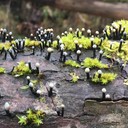
From my archives, here is an organism which I could not make a clue. I have only external images, no micro. I was thinking about some sort of structured mould (an anamorph of something maybe ?!?). Maybe someone has met something similar and can suggest some additional info... without expecting miracles! These were growing on fallen branches of Eucalyptus trees
Thanks in advance
Yes, I confirm that Cyanoboletus pulverulentus becomes deep blue in few seconds (=instantly). Problem with external colours form me is that they vary with maturation and also interspecific variety. Old specimens of Cyanoboletus are really dull and unattractive, young ones are quite more eye-pleasing. Thanks for your explanation Seb.
L.G.
Stephen
For my learning ... could the 6th mushroom which stains deep blue be Cyanoboletus pulverulentus? (also an oak lover!)
Thanks
Hello again. I found a few examples of this beautiful Lactarius with a burnt orange pileus and a reddish stipe which gradually deepens to blackish-red at the base. Spores subglobose with reticular ornamentation around 8 um diameter. Sap not abundant turbid (not milk-white) slightly bitter but not strongly so. I was thinking that this is the Lactarius atlanticus but after reading this site:
two look likes are mentioned: L. serifluus and L. subumbonatus, but I think both are not the same that I have photographed. The atlanticus epithet (hence indicating its distribution is far west) and the dark red stipe put some doubt in my mind... BUT I think I should not look elsewhere and confirm this as L. atlanticus!
Thank you.
Here Limacella is scarce-frequent and I think there is a single species. It is in the whereabouts of L. furnaceae or L. subfurnaceae, but I can't get the right knowledge how to differentiate between, if ever they are really different. I have plenty of material (exsiccata), images and micro-data to share if someone like Mollisia whish to investigate further. The ones I found are almost always associated with leaf/branch litter of carob trees. The smell is really distinct !
At this stage, I am sure that the genus is Pseudobaeospora (family Tricholomataceae) - hence that is already an equalizer![]()
Regarding the species, that is a challenging task, and I try to get as much info as possible, but I already pinned P. albidula and P. calcarea are at the moment the strongest options.
(PDF) Pseudobaeospora albidula (Agaricales) found in the Czech Republic
This paper has an interesting discussion opening to further research and options, from a possible new species that gros on leaf litter of broad leaf trees (spec. to carobs?) because from what I gather, these 2 species prefer to grow on moss.
Then this is also interesting:
Pseudobaeospora paulochroma and P. bavariae differ from P. albidula and P. calcarea in a positive (ie yellow or pale sordid yellowish) reaction of the pileus surface with KOH. Pseudobaeospora terrayi has a mild taste and therefore dif - fers by a pale yellowish greenish reaction of the pileus surface with KOH and by the presence of well-developed suprapellis.
(14) (PDF) Pseudobaeospora albidula (Agaricales) found in the Czech Republic . Available from: (PDF) Pseudobaeospora albidula (Agaricales) found in the Czech Republic [accessed Nov 03 2021].
I judge a positive pale sordid yellowish reaction in my specimen (see photo above)!
Some micro images (not much to show!)
good evening mycophiles
This fungus is the same that I reported weeks ago but here it is from a different location:
Small white mushroom, with a tomentose covering
These are the new images taken yesterday.
MACRO:
Small basidiocarps with a tomentose veil, hence tomentose cap and ring.
Gregarious or fused at the base
Lamellae free but very close to the stipe
Taste fungoid-sweet, good and palatable
Scent indistinct
Stipe white, but sometimes greyish esp. after handling
Pruinose-puberulent esp. above the ring
Stipe hollow, inside there are longitudinal white fibres (not sure if it is a characteristic of species)
Base slightly swollen in solitary examples
Ring slowly evanescent, leaving a cottony scar
Spores hyaline, don't stain easily.
4% KOH on stipe forms a faint yellowish colour (with a hint of greenish tones?)
MICRO (quite boring)
Cheilocystidia and pleurocystidia not observed
Distinct feature: Spores 2.0-3.0 um which aggregates in pairs or clusters, pip-shaped.
Basidia 4-spore (I managed to see one!)
Veil (or pileipellis) trichoderm of cylindrical hyphae, a few with clamp junctions, unspecialised
Steve vs White fungus with ring: 0-1 in 2018!
Now I met the same fungus in the same locality and even almost the same date (23/10/2018) and I want to rechallange and try to identify this cute and curious species. I just came from the forray (super tired!) and still have my camera in the bag, but these are the images from 2018. I will document gradually this fungus here. It is small (2 cm) and grow in small clusters, with the base of the stipe fused / connexed. I am quite sure that the habitat is leaf litter of Quercus ilex or Ceratonia siliqua, not connifer ( pine leaves present in pic). Investigation for tonight or tmrw!
To be continued...
Ah ok for the BWB ![]() Strange u have that violet tinge, but I have to say that it did not stain the spores very well, so better you buy a fresh supply. It is not expensive. IN my humble opinion it should stain both in acid and in water but it does not work well when the pH is slightly alkaline (unlike Congo red). In pH > 10 or so it will precipitate or form some blackish colour if my memory is still good. Well ... just buy a new BWB and see if u can compare the spores of the same sample
Strange u have that violet tinge, but I have to say that it did not stain the spores very well, so better you buy a fresh supply. It is not expensive. IN my humble opinion it should stain both in acid and in water but it does not work well when the pH is slightly alkaline (unlike Congo red). In pH > 10 or so it will precipitate or form some blackish colour if my memory is still good. Well ... just buy a new BWB and see if u can compare the spores of the same sample
Thank you Tuppie - enigma solved!!! I decrease the temp to 40C.
Good day!
Dear friends. Some years ago I built my own desiccatorfresh with temperature control, light, fan, etc. I wanted to ask if 42-49C is the ideal temp to desiccate fungi in a challenging climate with humidity >75% and warm winter temperatures where infections from moulding parasites are not uncommon. Is >50C to high that damages the hyphae/cystidia? TNX
P.s. I place the fresh fungi inside, and those dry I keep them on the lid while working with them which is pretty warm until I am ready and store them in boxes later on.